Computer vision
Polyp segmentation
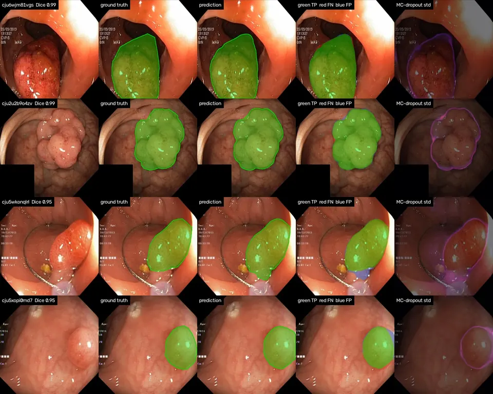
Colon polyp segmentation on Kvasir-SEG with MC-dropout uncertainty maps, checked against the model's own errors. A learning project, not for clinical use.
The problem
A segmentation mask on its own doesn't say when to trust it. Monte Carlo dropout gives a per-pixel uncertainty map almost for free, but it is often shown as a pretty heatmap and never checked. I trained a polyp segmenter on Kvasir-SEG and measured whether its uncertainty is actually high where it is wrong. It is a learning project, not a clinical tool.
What I built
- Split the 1,000 Kvasir-SEG frames 80/10/10 with a committed, seeded split, so the test set stays untouched.
- Trained a U-Net with an ImageNet ResNet-34 encoder on BCE plus soft Dice, with channel dropout after each decoder block, and kept the checkpoint with the best validation Dice.
- Kept dropout on at test time for 20 passes and scored the per-pixel standard deviation against the model's own errors (AUROC and Spearman).
- Exported to ONNX and CoreML, plus a second graph that runs the encoder once and the decoder 8 times, so the uncertainty map also runs in the browser.
Results
| Polyp size (share of frame) | Images | Mean Dice | Recall |
|---|---|---|---|
| Small (< 5 %) | 16 | 0.868 | 0.908 |
| Medium (5–20 %) | 51 | 0.938 | 0.937 |
| Large (> 20 %) | 33 | 0.867 | 0.843 |
| All | 100 | 0.904 | 0.901 |
What the results show
- 75 of 100 test images score above 0.9 and the median is 0.95. The mean is pulled down by four hard cases below 0.5, all flat or multi-part polyps that are only partly segmented.
- Errors are misses rather than false alarms: precision is 0.937 against recall 0.901.
- The uncertainty map finds boundary errors well. Its std is 0.103 on wrong pixels against 0.010 on correct ones, with a pixel-error AUROC of 0.930.
- It is a poor miss detector. The worst image ranks only 29th of 100 by uncertainty, because every dropout sample agrees the missed tissue is normal mucosa.
Limitations
- Single-centre data and my own split, so the numbers aren't strictly comparable to papers, and 100 test images leave about ±0.025 noise on mean Dice.
- Every Kvasir-SEG frame contains a polyp, so false positives on healthy tissue are unmeasured.
- MC dropout doesn't flag confident errors. A deep ensemble is the next comparison.
Full results, commands and figures are in the repository README.